Breast screening is an important part of looking after your health, but deciding which test is appropriate can sometimes be confusing. Mammograms and breast ultrasounds each have their unique advantages, and understanding how they work can help you and your doctor make an informed choice. Below is an overview of how these screening methods differ, what each one is suited to detect, and when one might be recommended over the other.
1. Mammogram: How It Works
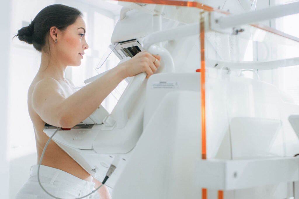
A mammogram is an X-ray of the breast. During the procedure, the breast is gently compressed between two plates to spread out the tissue, which allows for clearer images. This technique can reveal small lumps, tiny calcium deposits (known as microcalcifications), and structural changes that may not be detected through touch alone.
- Suitable for detecting:
- Early-stage growths or changes in breast tissue
- Microcalcifications that could indicate possible issues
- Considerations:
- Uses a low dose of radiation
- May cause brief discomfort due to compression
- Often recommended as a routine screening for women starting at a certain age, depending on individual risk factors
2. Breast Ultrasound: How It Works
A breast ultrasound uses high-frequency sound waves to produce images of the breast tissue. A gel is applied to the skin, and a handheld device called a transducer is moved over the area to capture real-time images. Ultrasound does not use radiation, which makes it a suitable option for younger individuals or those who may be pregnant or breastfeeding.
- Suitable for detecting:
- Fluid-filled cysts, which appear differently from solid lumps
- Areas of concern that need further evaluation after a mammogram
- Possible issues in individuals with dense breast tissue
- Considerations:
- Does not expose individuals to X-ray radiation
- Can help clarify whether a lump is solid or fluid-filled
- Sometimes used alongside a mammogram for a more detailed assessment
3. When Might You Need One Over the Other?
Mammogram First
Healthcare providers often recommend a mammogram as a starting point for routine breast screening, especially for women above a certain age or those with a family history of breast cancer. Mammograms are helpful in identifying early changes, such as microcalcifications.
Ultrasound for Further Assessment
If a mammogram identifies an area that needs more evaluation, or if there is a concern that might not be easily captured on a mammogram, an ultrasound may be advised. Ultrasound is also useful if you have dense breast tissue, which can sometimes make mammogram images harder to interpret. Additionally, pregnant or breastfeeding women may have an ultrasound to avoid exposure to any amount of radiation.
Combined Approach
In many cases, both tests are used together. A mammogram provides an overview of the breast, while an ultrasound offers a more targeted look at suspicious regions.
4. Making a Decision
Choosing between a mammogram and an ultrasound depends on several factors, including your age, medical history, breast density, and any symptoms you may be experiencing. It is important to speak with a medical professional who can recommend the most appropriate screening based on your individual circumstances.
Book a Consultation at The Radhika Breast & General Surgery Clinic
If you have concerns about your breast health or need guidance on whether a mammogram, breast ultrasound, or a combined approach is suitable for you, consider arranging an appointment with Dr Radhika Lakshmanan at The Radhika Breast & General Surgery Clinic. Dr Radhika Lakshmanan provides care that is tailored to each person’s needs, including screening advice, diagnosis, and treatment.
Taking a proactive step now can help you feel more at ease about your health. Book a consultation today and learn more about the screening method that may be right for you. Your well-being is our priority, and we look forward to supporting you on your path to a healthier future.